Migration Assay (Wound Healing Assay and Oris Assay)
Cell-Based Assays

Cell Migration Assays (Wound Healing Assay and Oris Assay)

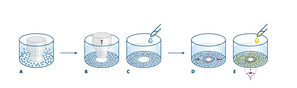
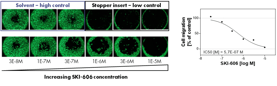
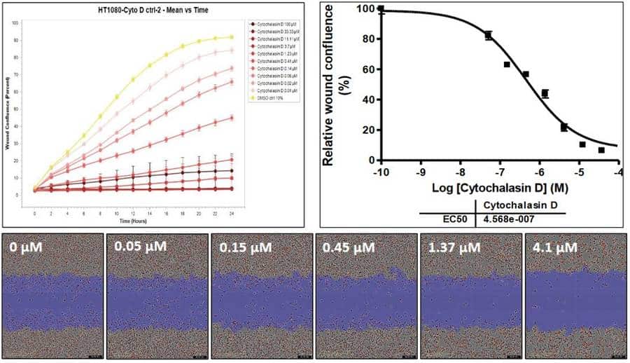
Cell migration assays are suited for the investigation of drug potency to reduce tumor cell metastasis. The migration of tumor cells is initiated by providing a gap in the cell monolayer that the cells close via undirected migration. The ORIS Assay is performed with a stopper for cells to grow around, whereas the Wound Healing Assay is based on a scratch that creates a gap in the cell layer.

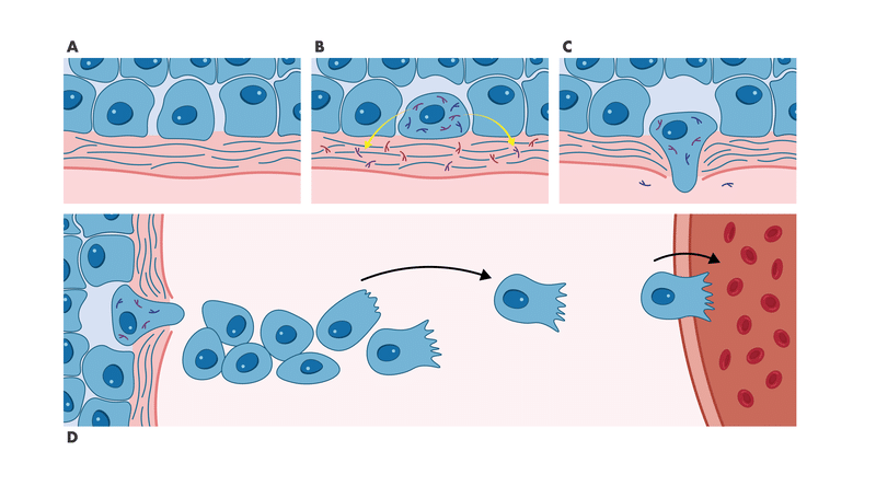
Cell migration is a hallmark in tumor development. It is relevant for angiogenesis to assure tumor nutrition as well as for the formation of metastases, in which tumor cells leave the primary tumor site and invade other tissues. The process of cell movement is induced by various agents such as growth factors and chemokines and is associated with complex signaling events that involve many components of the cellular motility machinery. Such signaling components (e.g. FAK, cSrc, ROCK), as well as ligand/receptor interactions that induce migration, represent attractive targets for tumor therapy.

The ORIS Assay and Wound Healing Assay may be used to characterize cell migration inhibitors and promoters. Both migration assays are valuable tools for academic researchers and pharmaceutical industries to determine the impact of new drug candidates on metastasis.

Reaction Biology’s tumor cell biology teams provide long-standing expertise in developing and running cell migration assays for standard compound screening and custom-tailored solutions. Get in contact today to discuss your research needs.

  • High-throughput compatible
  • Combination treatment is possible
  • Two cell migration assay formats are available: ORIS Assay and Wound Healing Assay (also called Scratch Assay)

Early Metastasis Process

The early steps of the metastasis process involve A) Loosening of the intercellular junctions between tumor cells, B) Degradation of the intracellular matrix of the basement membrane, C) Invasion into surrounding tissue, and D) Migration to endothelial vessels.

Steps A-C are modeled with the Invasion Assay. Step D is modeled with the Migration Assay.

tumor cell migration and invasion of tissue for metastasis

tumor cell migration and invasion of tissue for metastasis

Cell Migration Assay Details

  • ORIS Assay Description
  • ORIS Assay Example Data
  • ORIS Assay Details
  • Wound Healing Assay Description
  • Wound Healing Assay Example Data
  • Wound Healing Assay Details
ORIS Assay Description

ORIS Assay Description

The ORIS assay is well suited for the testing of compounds that inhibit undirected tumor cell migration. It is performed in a 96 well format and can be used for screening with medium throughput rates with high accuracy.

oris assay principle for testing compounds on tumor cells against migration

Assay procedure. (A) Tumor cells are seeded onto collagen I-coated multi-well plates equipped with a barrier that prevents cell adherence to the center of each well. After 18 hours, the barrier is removed (B) revealing a clear region in the center of the well. During a 24- to 48-hour incubation period in the presence of test compound (C), tumor cells migrate into this detection zone (D) which is monitored by fluorescent-labeled cells (E).

The assay is performed with the ORIS assay kit from Platypus.

ORIS Assay Example Data

ORIS Assay Example Data

oris assay example data are shown for reference compound ski-606 for screening of compounds inhibiting migration of tumor cells

ORIS Assay example data. A c-Src inhibitor was incubated with breast cancer cells for 48 hours, and the migration of cells into the detection zone was visualized by fluorescence photography (left), and results presented as a dose-response curve (right). Negative control = solvent; positive control = removal of the cell barrier at the end of the test compound incubation.

ORIS Assay Details

ORIS Assay Details

Setup: IC50 value determination. Drug combination testing is possible.

Controls: Removal of stoppers at the endpoint is the low control. DMSO only treatment is the high control. Reference control treatment is performed for quality control in every project.

Custom-tailored assays can be performed. This assay can be established with new cell lines according to the client’s research needs.

Turnaround time: Around 3 weeks. Expedited scheduling and data delivery can be arranged prior to study commencement.

Report: A detailed report including assay conditions, methodology, and comprehensive evaluation of data as well as raw data for own analysis will be provided. Photographs of colonies can be provided upon request.

Screening facility: The assay is performed in Freiburg, Germany.

Compound requirements: In brief, please provide at least 30 μl of a 1000x stock solution of the highest testing concentration in DMSO or the equivalent of solid powder.

Wound Healing Assay Description

Wound Healing Assay Description

The Wound Healing Assay (or Scratch Assay) measures the impact of compounds on undirected cell migration after wound-making via scratching the cell monolayer. The assay is suitable for investigation of tumor and normal cells and is performed with IncuCyte system which enables real-time monitoring of cell migration.

scratch assay procedure for compound testing on tumor cells for migration properties

Assay procedure. (A) Tumor cells grow to monolayers. (B) Using the WoundMaker, homogeneous scratches will be created mechanically with pins in a 96 well format. (C) After the application of the test compound, plates incubate in the IncuCyte S3 instrument for real-time quantification of tumor cells migrating into the scratch zone (D).

Wound Healing Assay Example Data

Wound Healing Assay Example Data

scratch assay example performed with incucyte for dose response studies for compound testing against migration of tumor cells

Effect of inhibition of cell migration on HT-1080 cells. HT-1080 cells with red fluorescence were seeded into ImageLock plates in complete growth media. After the cells reached about 90% confluence, wounds in all wells were simultaneously created using the WoundMaker tool. Cytochalasin D was added at serial dilutions and plates were placed in the IncuCyte instrument for scheduled scanning.

Upper left: Time course of the effect of an inhibitor of actin polymerization on wound healing over 24-hours. Upper right: Dose-dependent effect of the drug on wound healing at 8h time point. Lower panel: Representative images of wound healing at 8h time point. The blue areas represent the initial wound scratches.

Wound Healing Assay Details

Wound Healing Assay Details

Setup: IC50 value determination with 10 concentrations in triplicates performed in 96 well format. Drug combination testing is possible.

Controls: DMSO only control serves as high control. A reference compound recommended by the client is used for positive control.

Assay development: Custom-tailored assays can be performed. This assay can be established with new cell lines according to the client’s research needs.

Turnaround time: Around 3 weeks.

Report: Raw data, IC50 curves, and IC50 values as well as representative images of wound closure are provided in the report.

Screening facility: The assay is performed in Malvern, US.

Compound requirements: In brief, please provide 20-50 μl of 10-50 mM DMSO stock or the equivalent of solid powder. Please refer to our FAQs for information regarding compound preparation and shipping.