SubQperior Tumor Models
In Vivo Pharmacology

SubQperior™ Tumor Models

Standard implantation via subcutaneous injection causes frequent tumor ulceration resulting in early abrogation of studies, leaving researchers with too short treatment windows and high tumor heterogeneity with a poor statistical value of tumor study results. To overcome these challenges, we have developed a superior implantation method for syngeneic cell line-derived tumor models. SubQperiorTM: Tumor cell implantation via mammary fat pad injection.

  • SubQperiorTM implantation results in larger tumors and longer treatment windows
  • Homogeneous and reliable growth yields study outcomes with outstanding statistical value
  • Accelerate your drug discovery with the SubQperior Panel Screen for efficacy testing on 6 tumor models in one project.

SubQperior

We have developed an innovative approach for tumor cell implantation.

Our syngeneic subQperiorTM mouse models are based on mammary fat pad injection of tumor cells to overcome frequent challenges caused by tumor ulceration and resulting in homogenous tumor growth and thus in vivo studies with excellent statistical value.

undefined

Browse our subQperior tumor models

SubQperior In Vivo Studies

  • CT26wt Study Example
  • Standard Study Layouts
  • Study Options
  • Custom Model Development
  • Certification
  • Quality Assurance
  • Animal Welfare
CT26wt Study Example

Goal: The frequently used immune checkpoint inhibitors anti-mPD-1 and anti-mCTLA-4 were tested for their potential to inhibit subQperiorTM CT26wt primary tumor growth.

Study layout: CT26wt cells were implanted into the mammary fat pad of Balb/C mice, at 12 mice per group. After randomization on day 5, treatments were initiated on the same day. Test compounds anti-mPD-1 (Group 2) and anti-mCTLA-4 (Group 3), both administered i.p. at 10.0 mg/kg on days 5, 8, and 12, were evaluated versus the corresponding vehicle control (Group 1). Tumor growth was monitored by calipering twice per week, animal weight was measured three times per week, animal behavior was observed daily. On day 21, animals were euthanized and endpoint measurements for the determination of primary tumor volumes, and wet weights were performed.

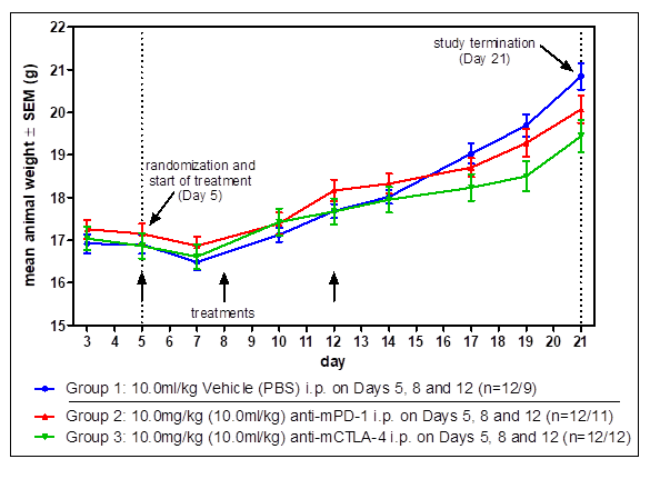
animal weight of CT26 tumor model study in mice

Animal weight of CT26wt subQperior tumor model is shown with vehicle and anti-mPD-1 and anti-mCTLA-4 treatment.

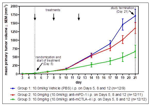
CT26 tumor growth with and without treatment for compound testing

Monitoring of CT26wt tumor growth was performed twice weekly via calipering. Shown are the mean values of tumor size.

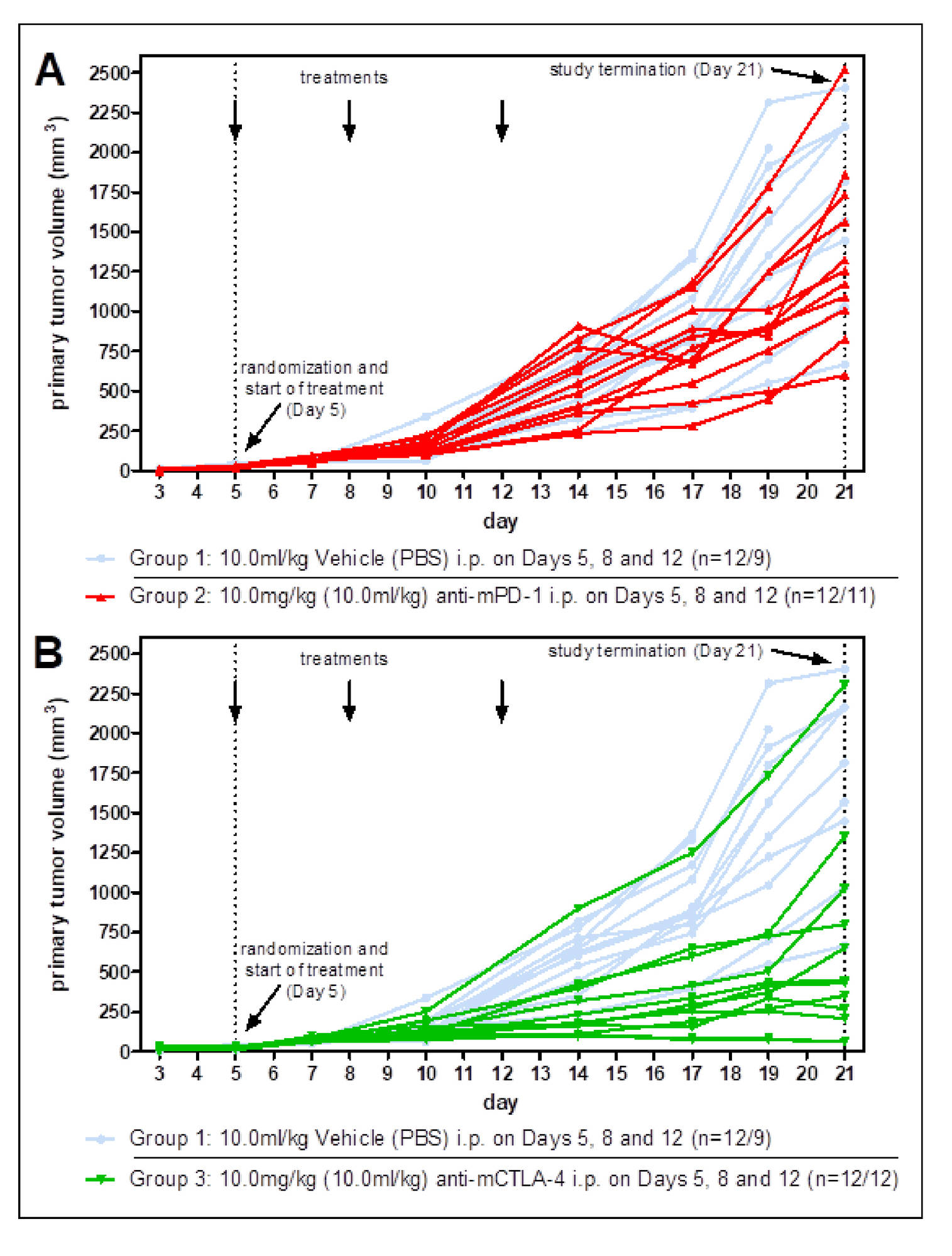
Tumor growth

Tumor growth is shown for individual tumors. upper blot: anti-mPD-1 treatment vs vehicle treatment. lower blot: anti-mCtLA-4 treatment vs vehicle treatment.

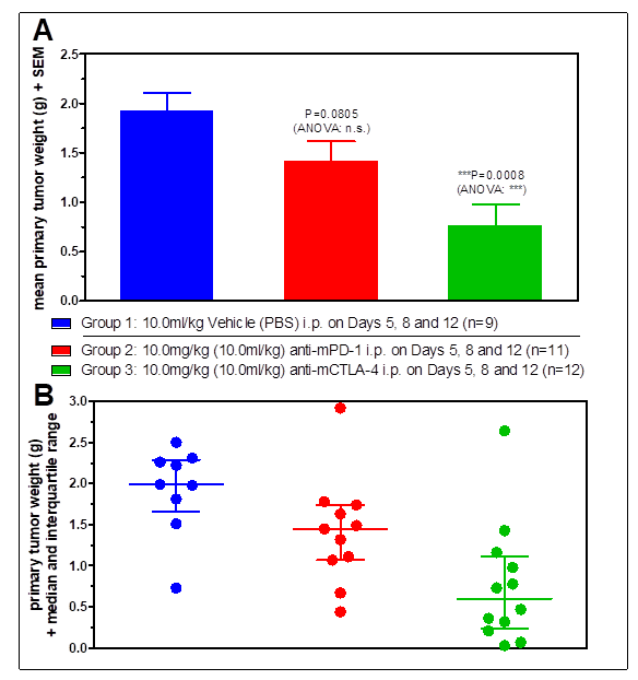
tumor sizes of CT26 tumors at termination as tumor weight and tumor volume

At day 21, tumors were isolated from the mice at necropsy and weighed. Blotted are the mean tumor weights in the top graph and the individual tumor weights in the lower graph together with their median values and interquartile ranges. P values were calculated compared to the vehicle control using the unpaired t‑test and the one-way ANOVA with Dunnett´s test.

Standard Study Layouts

Standard efficacy study

SubQperiorTM tumor models are monitored via calipering after tumor cells were implanted into the flank of the mice. When tumors reach the predetermined tumor volume, animals are randomized in treatment groups based on tumor size and treatment starts. The mice are monitored on a regular basis: tumor size is measured via calipering twice weekly, animal weights are taken three times per week and animal behavior is observed daily. Once per week, we update the customer with a graphical presentation of the study progress.

Report

A comprehensive report will be prepared by a PhD-level medical writer custom-tailored for each project with data that can be used for filing official documents. The report includes material, methods, raw data, animal health chart, and graphs plus statistical evaluation.

Selection of optional services:

  • Isolation of tumors at necropsy for determination of volume and wet weight
  • Preservation of tissue via fixation or snap-freezing
  • Histological or pathological analysis of tumor tissue and mouse body
  • Determination of expression of genes or proteins
  • Plasma sampling and bioanalysis of compound or metabolites
  • Quantification of cytokines and chemokines in blood or tissue via MESO QuickPlex SQ120 multiplex analysis
  • Flow cytometry analysis for immune cell profiling
Study Options

Standard Efficacy Studies: Determination of the therapeutic effect of a new drug on a syngeneic tumor model.

Panel Screening: On a regular basis, Reaction Biology performs panel screenings for the determination of therapeutic efficacy on 6 syngeneic tumor models. This economic screening option includes vehicle as well as anti-PD1 treatment arms – free of charge.

Proof-of-Concept Studies: New drug candidates need to be tested not only for efficacy but also for their mechanism of action in the animal. For example, exploratory proof of concept studies can be performed via flow cytometric analysis for investigation of effects on tumor-infiltrating immune cells.

Pharmacokinetic Studies: Pharmacokinetic studies on tumor-bearing mice are helpful in determining the concentration of drugs not just in plasma but in the tissue of the tumors.

Pharmacodynamic Studies: Pharmacodynamic studies elucidate whether and how a drug acts on its target for example, via flow cytometric analysis of tumor-infiltrating immune cells.

Dose-Response-Relationship: Determine suitable drug doses for efficacy testing.

Drug Combination Studies: Combinations of drugs can lead to synergistic effects and vastly increased tumor response. Generally, we validate our syngeneic tumor models with immune-checkpoint inhibitors anti-PD1, anti-PDL1, and anti-CTLA4, which are often used for combination therapy.

Survival Studies: A survival study setup is used to obtain information about a long-term effect of the test substance in comparison to control. Survival studies give meaningful results for ambivalent drug candidates measuring the number of responders and non-responders.

Custom Model Development

We continuously increase our portfolio of tumor models and provide custom development of new tumor models. Ask us for cost-sharing options.

Cell lines: We can develop new models based on customer-provided cell lines or purchase a cell line from a commercial vendor.

Establishment study: We will monitor the tumor growth after tumor cell implantation into 12 mice of the mouse strain matching the tumor cell‘s origin for the determination of the tumor growth characteristics.

Immune-checkpoint inhibitors: Most of our models were tested with a variety of immune-checkpoint inhibitors, which can be used for combination drug studies.

Certification

The animal facility of Reaction Biology is located in Freiburg, Germany.

Our facility is certified under ISO 9001:2015, which is an international standard that specifies requirements for the quality management system and demonstrates the ability to consistently provide products and services that meet customer and regulatory requirements. Reaction Biology is committed to continuously maintain and improve its quality management system as a key element for the achievement of the highest customer satisfaction. ISO 9001
Quality Assurance

Cell lines

  • Routine authentication of tumor cell lines by STR profiling
  • Mycoplasma testing of tumor cells by PCR just prior to implantation

Animal work

  • Routine health monitoring of sentinel animals (according to FELASA guidelines)
  • Standardized operation procedures are in place of every step and every model

Study support

  • Studies generally start (tumor implantation) 3 to 5 weeks after receipt of the order
  • Suggestions for study layout to get statistically relevant results
  • Weekly study update and personal contact with the study supervisor
  • Reports are written by medical writers on Ph.D. level

Ethical principles

  • Our animal work is conducted according to the 3R (Replacement, Reduction, and Refinement)
  • We are working closely with our animal care committee to ensure timely adaptions of our animal care licenses to custom-tailor our client’s project to provide the most meaningful study outcome
Animal Welfare

Reaction Biology uses laboratory animals to help our customers to understand the fundamental mechanisms behind malignancies and to discover therapeutics to prevent and treat cancer. Data obtained from animal models is critical to predicting the clinical outcome for an oncology drug candidate in development.

Animal welfare is of the utmost importance to us. Animal-based research is highly regulated to ensure ethical and responsible treatment. The mice in our facility are specifically bred for research purposes, and they are cared for to the very high standards.

We are working under GV-SOLAS and ISO9001 regulations in regard to standards of animal welfare and code of practice.

We employ three veterinarians and appropriately trained staff to ensure animal welfare is maintained at the highest standards. Regulation officers inspect our unit regularly.