Flow Cytometry Applications
Biomarker Discovery

Flow Cytometry Applications

Flow cytometry is a versatile tool that efficiently allows the investigation of cells after compound treatment based on changes in protein expression, protein localization, or protein modifications. For example, flow cytometry is used to quantify the activity of drug candidates via quantification of substrate modifications, including phosphorylation. Other cellular processes such as apoptosis or cell signaling can also be measured.

At Reaction Biology, flow cytometry can be conducted with cell culture, primary cells, tissue material, or blood. Our scientists have access to a full suite of state-of-the-art flow cytometers and cell sorters and can further analyze sorted cells via qPCR, immunofluorescence, or other readouts.

Our services include:

  • Single-plex or multi-plex capacity with up to 20 markers
  • Quantification of drug treatment effects on a cellular level
  • Custom-tailored project design and assay development
  • Fresh samples can be created in-house by our cell assay or animal testing teams

Information about flow cytometry of tumor material for immunophenotyping can be found here.

Connect with our scientists to find out how we can help you achieve your research goals and determine which methods are best suited to monitor the effects of your drug candidate.

Instrumentation – Flow cytometers and Cell sorters

Examples of Flow Cytometry Applications

  • Target expression in tumor material
  • Cell division via CFSE staining
  • Intracellular protein expression
  • Immunophenotyping
Target expression in tumor material

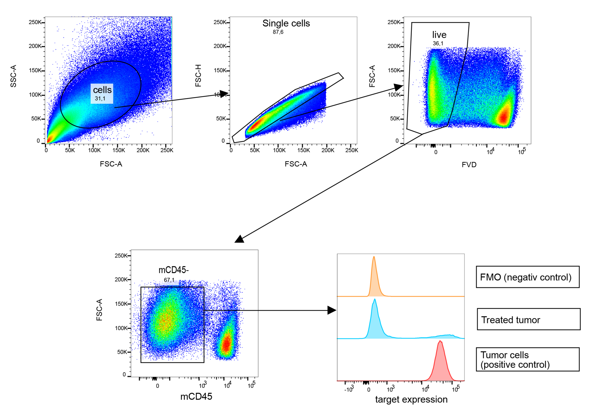
Example: Investigating the expression level of the target protein after tumor treatment

Human JIMT-1 breast cancer tumors were grown in mice treated with vehicle or test substance. At study endpoint, tumors were isolated, and tumor material was disrupted with gentleMACS C tube containers and the Tumor Dissociation Kit (Miltenyi Biotec) before antibody staining and flow cytometry analysis.

Gating: Single cells were selected for live events via live/dead dye. Human tumor cells were identified as negative for mouse CD45 marker and investigated for target expression on the cell surface. Untreated tumor cells served as positive control expressing the target on 99% of cells, whereas only 14% of the tumor cells from treated animals were stained positive for the target.

In conclusion, treatment of JIMT-1 breast cancer with the test agent reduced the number of target expression tumor cells to 14%.

Cell division via CFSE staining

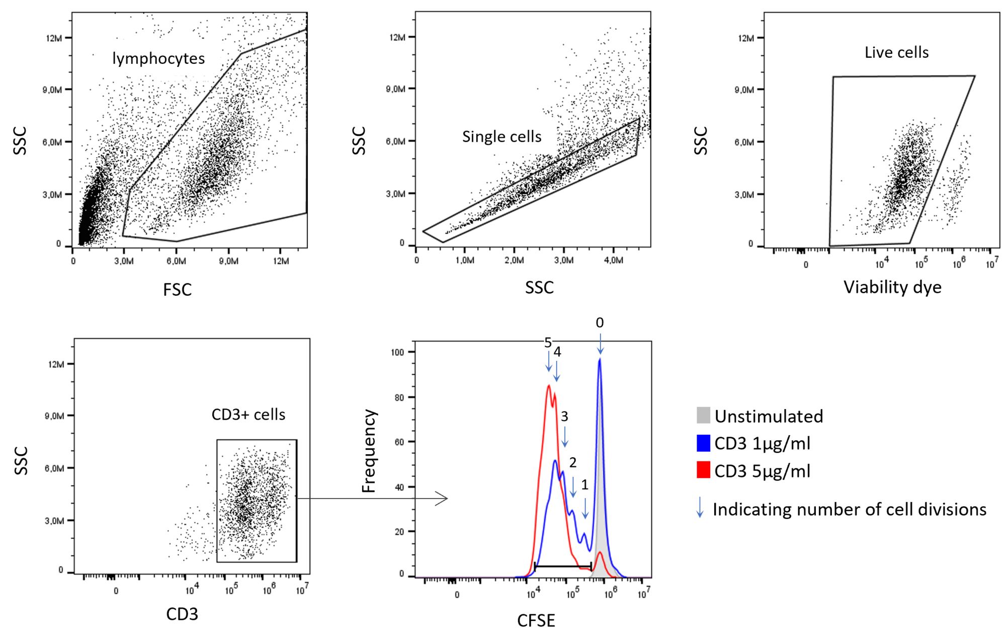
Example: Tracking cell division events by measuring the fluorescent dye CFSE in proliferating cells

CFSE (Carboxyfluorescein succinimidyl ester) is a cell-permeable dye covalently binding to amides creating fluorescent dye-protein conjugates. The dye is very stable and can be tracked for several days. The dye is still present in the daughter cells after cell division, each cell carrying half of the CFSE molecules, resulting in cell populations with varying degrees of fluorescence as cells continue to divide. Therefore, the dye is suitable for tracking cell division events.

In this example study, we labeled human peripheral blood mononuclear cells, also called PBMCs, with CFSE in the presence of two concentrations of plate-bound CD3 which triggers the proliferation of CD3-positive T cells. After 5 days, the cells were fixed with paraformaldehyde and measured by a flow cytometer.

Intracellular protein expression

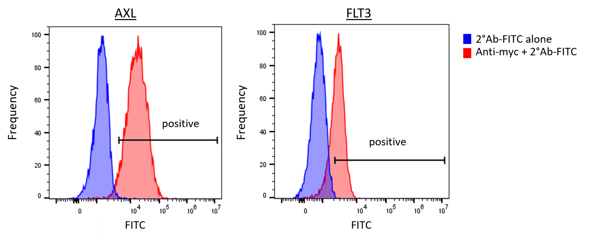
Example: Quantification of cells expressing an intracellular protein via the Tet-off system.

Mouse embryo fibroblasts (MEF) expressing the myc-tagged proteins AXL and FLT3 with a Tet-off system were fixed and permeabilized with ice-cold methanol. The cells were rehydrated with PBS and 1% fetal calf serum and stained with the primary antibody against myc, followed by a secondary anti-mouse antibody. Fluorescence was measured with a flow cytometer.

Immunophenotyping

Investigating the immune-modulatory action of drugs can be performed at the endpoint of an animal study at Reaction Biology. Flow cytometry is one tool to examine immune cell polarization and frequencies. Information about flow cytometry of tumor material for immunophenotyping can be found at the link here.